7.2: Kingdom Monera
Biology → Biology → CLASSIFICATION → CLASSIFICATION → Five Kingdom Classification | Author: admin | Feb 24, 2026
Kingdom Monera: Overview
Kingdom Monera includes all prokaryotic organisms, primarily bacteria. These are the most ancient, abundant, and widespread microorganisms on Earth. They are unicellular, lack a true nucleus (prokaryotic), and show remarkable adaptability, occurring in soil, water, air, extreme environments (hot springs, deserts, deep oceans, snow), and as parasites or symbionts in other organisms. Bacteria play crucial roles in ecosystems as decomposers, nitrogen fixers, and producers (in case of cyanobacteria). In Whittaker's five-kingdom system, Monera is distinguished by its prokaryotic nature, contrasting with eukaryotic kingdoms.
Key general characteristics:
- Unicellular and microscopic (typically 0.1–10 μm).
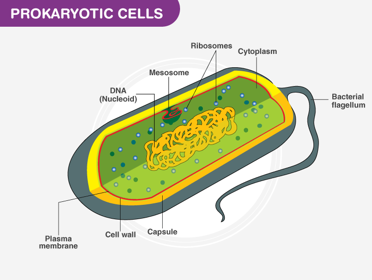
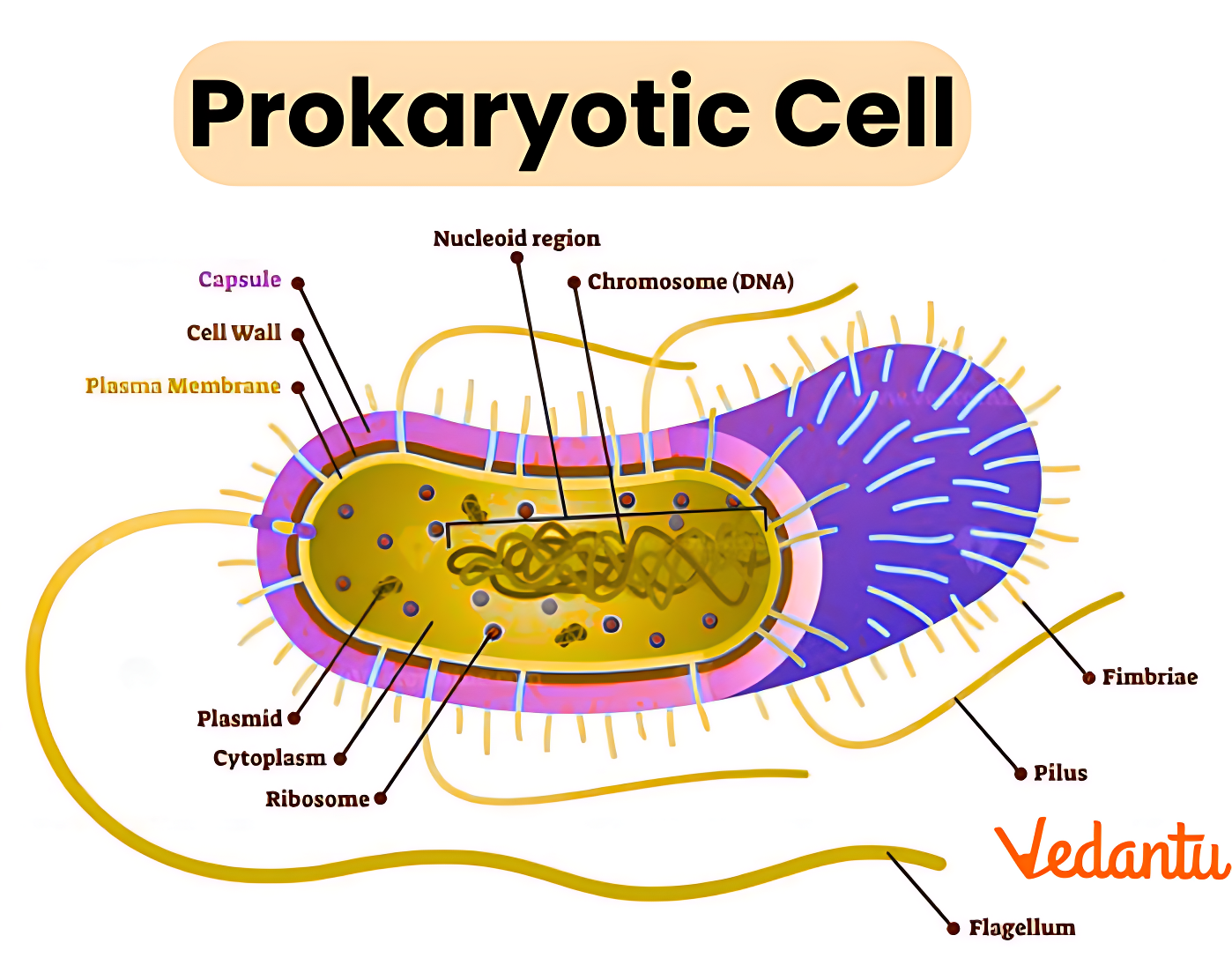
- Prokaryotic: No membrane-bound nucleus (DNA in nucleoid region), no organelles like mitochondria, ER, Golgi, plastids, or lysosomes.
- 70S ribosomes for protein synthesis.
- Cell wall present in most (made of peptidoglycan/murein, non-cellulosic); absent in Mycoplasma.
- Nutrition: Autotrophic (photosynthetic or chemosynthetic) or heterotrophic (saprophytic, parasitic).
- Reproduction: Primarily asexual (binary fission); under stress, spore formation; genetic exchange via conjugation, transformation, transduction.
- Locomotion: Flagella (if present), gliding, or none.
- Respiration: Aerobic, anaerobic, or facultative.
Nuance: While mostly unicellular, some form colonies or filaments (e.g., cyanobacteria), but lack true multicellular organization with tissue differentiation. Implications: Their simplicity enables rapid reproduction (some divide every 20 minutes) and high adaptability, making them vital for biogeochemical cycles but also pathogenic.

Shapes of Bacteria
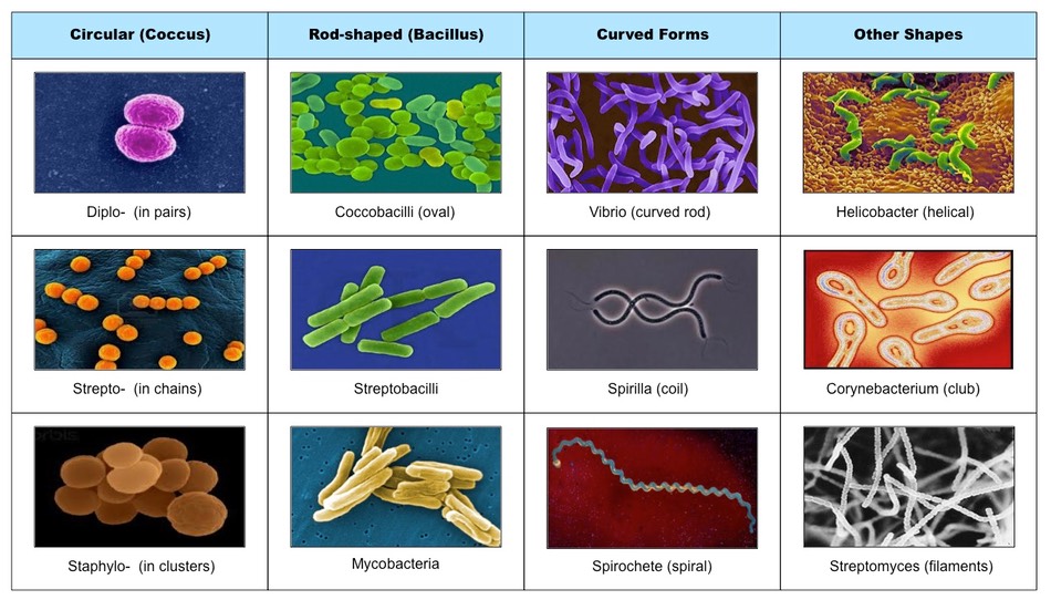
Bacteria are classified by shape, aiding identification and understanding adaptations (e.g., spirals for motility in viscous environments).
- Coccus (spherical): Single (monococcus), pairs (diplococcus), chains (streptococcus), clusters (staphylococcus).
- Bacillus (rod-shaped): Single, chains (streptobacillus).
- Vibrio (comma-shaped): Curved rods, e.g., Vibrio cholerae.
- Spirillum (spiral): Rigid spirals with flagella at ends.
Edge case: Pleomorphic bacteria change shape (e.g., some under stress). Related: Arrangement influences virulence (clusters resist phagocytosis).


Subgroups in Kingdom Monera
Monera divides into Archaebacteria (ancient, extremophiles) and Eubacteria (true bacteria, including cyanobacteria).
- Archaebacteria: Differ from eubacteria in cell wall (no peptidoglycan), membrane lipids, and rRNA. Thrive in extreme conditions (halophiles: high salt; thermophiles: high temperature; methanogens: produce methane in anaerobic environments like swamps, guts).
- Implications: Suggest early life evolution; methanogens contribute to greenhouse gases.


- Eubacteria: True bacteria with peptidoglycan wall. Include:
- Photosynthetic autotrophs: Cyanobacteria (blue-green algae) – oxygenic photosynthesis, chlorophyll a, phycobilins; form stromatolites, fix nitrogen.
- Chemosynthetic autotrophs: Nitrifying bacteria (Nitrosomonas, Nitrobacter), sulfur bacteria.
- Heterotrophs: Saprophytic (decomposers), parasitic (pathogens like Mycobacterium tuberculosis).
Special Features
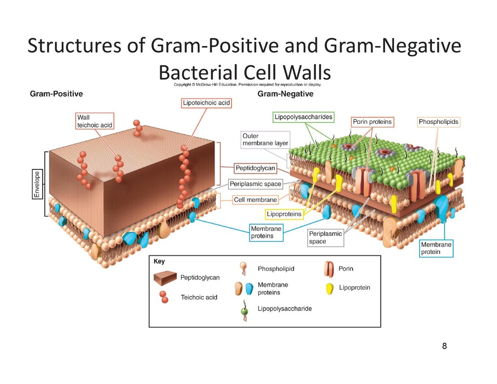
- Cell Wall and Gram Staining: Peptidoglycan layer; Gram-positive (thick peptidoglycan, purple stain) vs Gram-negative (thin peptidoglycan + outer membrane with lipopolysaccharides, pink stain). Affects antibiotic resistance and pathogenicity.
- Economic/Ecological Importance:
- Beneficial: Nitrogen fixation (Rhizobium in legumes), curd/yogurt (Lactobacillus), antibiotics (Streptomyces), bioremediation.
- Harmful: Diseases (typhoid, cholera, tuberculosis), food spoilage.
- Edge case: Mycoplasma – wall-less, smallest, cause pneumonia.
Overall, Monera's diversity and roles highlight prokaryotes as foundational to life, with implications for biotechnology, medicine, and climate.
Section 2: Quick Revision, Tips, Traps
Quick Revision
| Feature | Details | Examples/Notes |
|---|---|---|
| Cell Type | Prokaryotic, unicellular, no true nucleus (nucleoid) | All Monera |
| Cell Wall | Peptidoglycan (most); absent in Mycoplasma | Gram +ve thick; Gram -ve thin + LPS |
| Shapes | Coccus, Bacillus, Vibrio, Spirillum | Streptococcus (chain coccus) |
| Subgroups | Archaebacteria (extremophiles); Eubacteria (true bacteria, cyanobacteria) | Methanogens; Rhizobium |
| Nutrition | Autotrophic (photo/chemosynthetic), Heterotrophic (saprophytic/parasitic) | Cyanobacteria (photo), Nitrosomonas (chemo) |
| Reproduction | Binary fission, spores, conjugation | Rapid under favorable conditions |
| Special | Nitrogen fixation, oxygen production by cyanobacteria | Rhizobium in root nodules |
- Key Mnemonics: Shapes – "Cows Bite Very Spicy" (Coccus, Bacillus, Vibrio, Spirillum). Subgroups – "Ancient Extreme Bacteria" (Archaebacteria) vs "Everyday Usual Bacteria" (Eubacteria).
- Important Points: Only prokaryotes in five kingdoms; cyanobacteria as "blue-green algae" but bacteria; no multicellular true forms.
Exam Tips
- Draw and label prokaryotic cell, bacterial shapes, Gram wall differences – frequent in descriptive answers (SSC/RRB/JE).
- Link to ecology: Nitrogen cycle (fixers), decomposition, oxygen release (cyanobacteria) – UPSC/SSC favor application-based questions.
- Use examples: Rhizobium (beneficial), Salmonella (pathogenic).
- Compare Archaea vs Eubacteria: Cell wall, habitat – emerging in TGPSC/UPSC.
- MCQs: Focus on "which is prokaryotic?", "who fixes N2?", "Gram staining basis".
Traps to Avoid
- Don't call cyanobacteria "algae" – they are bacteria (Monera), not Plantae/Protista.
- Trap: Mycoplasma has no cell wall – not all Monera have walls.
- Common error: Archaebacteria are not "ancient bacteria" in outdated sense; they are distinct domain now, but NCERT places in Monera.
- Avoid confusing Gram +ve/-ve with shapes or nutrition.
- Nuance trap: All Monera unicellular – no true tissues; colonies don't count as multicellular.
- For exams: Viruses not in Monera (acellular).
Section 3: Previous Asked Questions-Answers and Expected Questions-Answers
Previous Asked Questions-Answers
(From UPSC, SSC, RRB, JE, TGPSC/TGLPRB papers, 2018–2025 compilations):
- Bacteria belong to which kingdom in Whittaker's classification? (SSC CGL 2020, RRB NTPC 2021) Answer: Kingdom Monera (all prokaryotes).
- Which of the following is prokaryotic? (UPSC Prelims 2019, TGPSC 2023) Answer: Bacteria (Monera); options often include amoeba (Protista), fungi.
- Cyanobacteria are included in which kingdom? (SSC CHSL 2022, RRB JE 2020) Answer: Monera (photosynthetic prokaryotes, formerly blue-green algae).
- The cell wall of bacteria is made up of? (SSC MTS 2018, TGLPRB 2022) Answer: Peptidoglycan (murein).
- Which bacteria fix atmospheric nitrogen in leguminous plants? (UPSC 2021, TGPSC Group 2 2024) Answer: Rhizobium (symbiotic in root nodules).
- Archaebacteria differ from eubacteria in? (SSC CGL 2023) Answer: Cell wall composition (no peptidoglycan), membrane lipids, extreme habitats.
- Which is absent in prokaryotic cells? (RRB Group D 2019, JE 2021) Answer: Membrane-bound nucleus/organelles.
- Shapes of bacteria include? (SSC 2024) Answer: Coccus, bacillus, vibrio, spirillum.
- Mycoplasma lacks? (TGPSC 2025) Answer: Cell wall.
- Gram-positive bacteria stain? (UPSC 2022) Answer: Purple (thick peptidoglycan).
Expected Questions-Answers
(For 2026 exams – UPSC, SSC, RRB, JE, TGLPRB, TGPSC):
- Describe the structure of a typical bacterial cell. (Expected UPSC Mains/SSC descriptive) Answer: Prokaryotic with cell wall (peptidoglycan), plasma membrane, nucleoid (circular DNA), 70S ribosomes, cytoplasm, possible flagella/pili/capsule; no membrane organelles.
- Differentiate Archaebacteria and Eubacteria. (Expected SSC CGL 2026) Answer: Archaebacteria: No peptidoglycan, branched lipids, extremophiles (e.g., methanogens); Eubacteria: Peptidoglycan wall, straight lipids, widespread (e.g., cyanobacteria, pathogens).
- Role of cyanobacteria in environment? (Expected RRB JE 2026) Answer: Photosynthetic, produce oxygen, fix nitrogen, form blooms; ancient oxygen contributors.
- Why is Gram staining important? (Expected TGPSC 2026) Answer: Differentiates bacteria (Gram +ve thick wall, antibiotics like penicillin effective; Gram -ve outer membrane, more resistant).
- Give examples of beneficial and harmful bacteria. (Expected TGLPRB 2026) Answer: Beneficial: Lactobacillus (curd), Rhizobium (N2 fixation); Harmful: Mycobacterium (TB), Vibrio cholerae (cholera).
- Which Monera member has no cell wall? (Expected UPSC Prelims 2026) Answer: Mycoplasma.
- Explain binary fission in bacteria. (Expected SSC CHSL 2026) Answer: Asexual: DNA replicates, cell elongates, septum forms, divides into two identical daughters; rapid under ideal conditions.
- How do archaebacteria survive extremes? (Expected RRB NTPC 2026) Answer: Unique membrane lipids, enzymes stable at high temp/salt/acid; e.g., thermophiles in hot springs.
- Difference between prokaryotic and eukaryotic cells (focus Monera). (Expected JE 2026) Answer: Prokaryotic (Monera): No nucleus/organelles, 70S ribosomes, peptidoglycan wall; Eukaryotic: Nucleus, organelles, 80S ribosomes.
- Importance of bacteria in nitrogen cycle? (Expected TGPSC 2026) Answer: Fix N2 (free-living Azotobacter, symbiotic Rhizobium), nitrification (Nitrosomonas), denitrification.